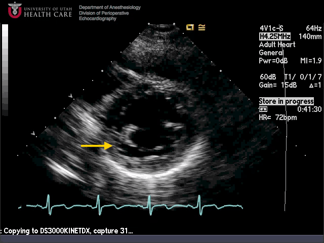
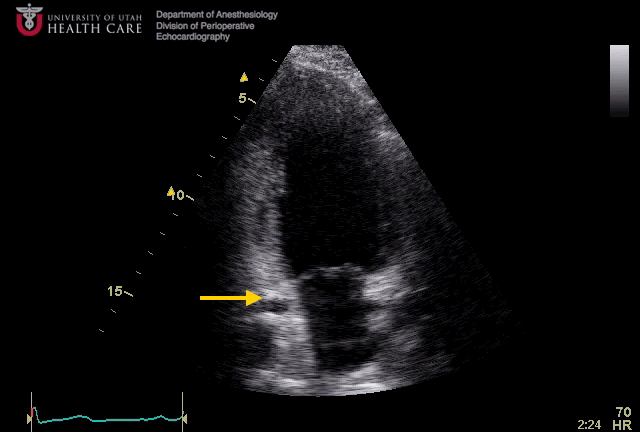
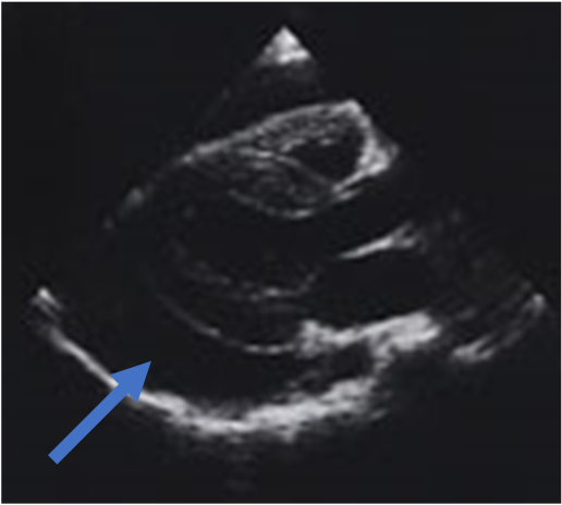
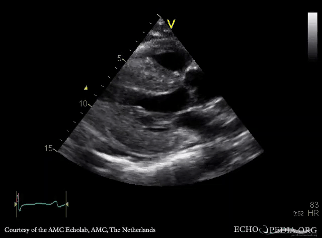
What view is this and what is the arrow pointing at?
![]()
- Parasternal LV short axis, basal inferior segment
- Subcostal LV short axis, basal inferior segment
- Parasternal LV short axis, basal anterior segment
- Subcostal LV short axis, mid inferolateral segment
- Parasternal LV short axis, mid anterior segment
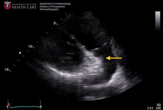
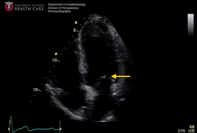
What view is this and what structure is shown by the arrow?
![]()
- Apical 4 chamber, left atrium
- Parasternal RV outflow, main PA
- Parasternal RV inflow, right atrium
- Apical 2 chamber, left atrium
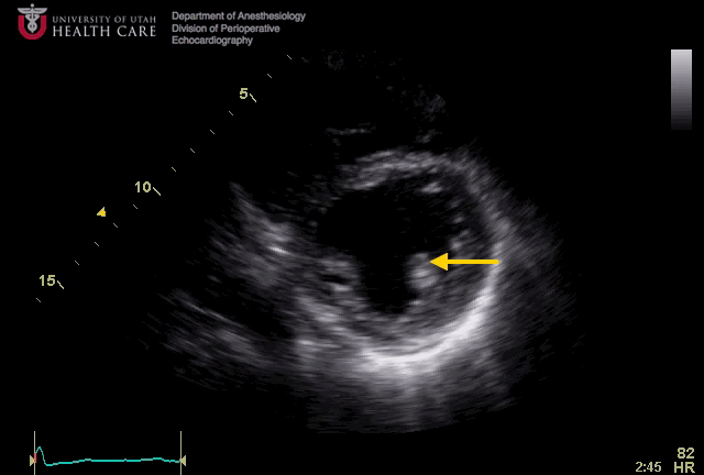
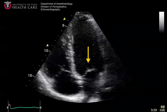
What view is this and what is the arrow pointing at?
![]()
- Transgastric LV short axis, posteromedial papillary muscle
- Parasternal LV short axis, posteromedial papillary muscle
- Parasternal LV short axis, anterolateral papillary muscle
- Transgastric LV short axis, anterolateral papillary muscle
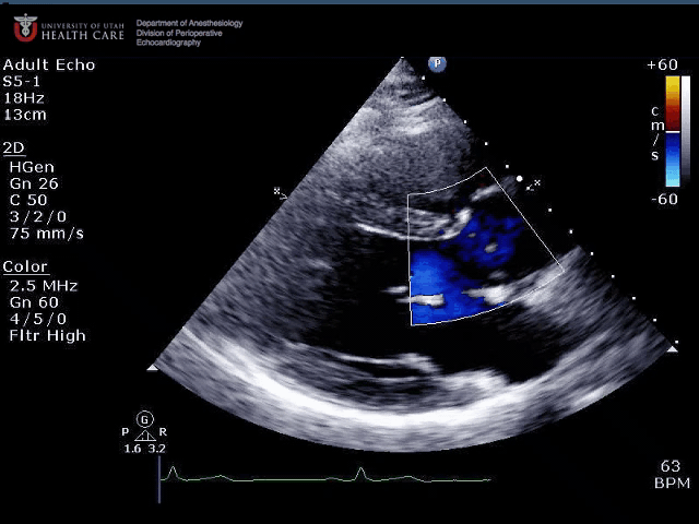
What view is this and what is seen?
![]()
- Parasternal long axis, color flow across the aortic valve concerning for stenosis
- Apical long axis, color flow across the aortic valve concerning for dynamic outflow obstruction
- Parasternal long axis, normal color flow across the aortic valve
- Parasternal long axis, color flow across the aortic valve concerning for insufficiency
What view is this and what structure is shown by the arrow?
![]()
- Apical 4 chamber, right atrium
- Parasternal long axis, aorta
- Apical long axis, right atrium
- Apical 2 chamber, left atrial appendage
- Apical 2 chamber, coronary sinus
What view is this and what structure is indicated?
![]()
- Apical long axis, anterior mitral leaflet
- Apical 4 chamber, septal leaflet of tricuspid valve
- Apical long axis, aortic valve
- Apical 4 chamber, posterior mitral leaflet
- Apical 4 chamber, anterior mitral leaflet
What view is this and what structure is indicated by the arrow?
![]()
- Subcostal 4 chamber, posterior mitral leaflet
- Subcostal 4 chamber, anterior mitral leaflet
- Apical 4 chamber, anterior mitral leaflet
- Apical 3 chamber, aortic valve
- Apical 4 chamber, posterior mitral leaflet
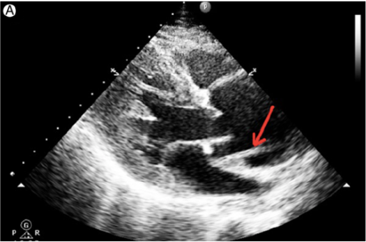
What view is this and what is shown by the arrow?
![]()
- Parasternal Long Axis
- Parasternal Short Axis
- Aortic Dissection
- Vegetation
- Coronary Sinus
Parasternal long axis, Dissection flap
This patient is likely to have?
![]()
- Severe aortic stenosis (AS)
- Severe mitral regurgitation (MR)
- Severe pulmonary hypertension
- Mild AS
What does the arrow show?
![]()
- Pericardial effusion
- Pleural Effusion
Optimisation of an image is essential when scanning which of the following statements are true:
- Increasing the depth will increase the frame rate
- Decreasing the sector width will decrease frame rate
- Focussing cannot be controlled
- Image quality is affected by frame rate
- The higher the scanning frequency, the better the resolution
Characteristic features of Amyloid Heart include:
- Bi-ventricular hypertrophy 双室肥厚
- Normal systolic function 收缩功能正常
- Mitral Valve thickening 二尖瓣增厚
- Intraventricular septal width greater than 2.0cm 室间隔宽度大于 2.0cm
- Normal left ventricular cavity size 左室大小正常
Aortic Regurgitation( Which are true? )
- Is a recognised complication of infective endocarditis 是公认的感染性心内膜炎并发症
- Is always severe if there is flow reversal in the aortic arch 如果主动脉弓内有血流逆转,总是很严重的
- With a continuous wave pressure half time of 350 ms is moderate 连续波压力减半时间为 350 ms 是中等反流
- With a colour flow map to LVOT height ratio of 50% is severe 彩色血流图与 LVOT 高度比为 50% 是重度的
- Can be underestimated when LVEOP is elevated 当 LVEOP 升高时可能会被低估
Aortic Valve Stenosis (Which are true?)
- Is most commonly secondary to Rheumatic Fever 最常见的是继发于风湿热
- Pressure gradient approximates to the square of the blood flow velocity across the valve 跨瓣压差近似于整个瓣膜的血流速度的平方
- Can cause dilatation of the ascending aorta 会导致升主动脉扩张
- Is assessed using the continuity equation 使用连续性方程进行评估
- Causes left ventricular dilatation early in the disease process 在疾病过程的早期引起左心室扩张
The following measurements are made in a patient. LVOT diameter=2.0cm, transaortic peak velocity=3.2m/s, left ventricular outflow tract peak velocity=0.9m/s, left ventricular EF=35% (Which are true?)
对患者进行以下测量。LVOT 直径 = 2.0cm,经主动脉峰值速度 = 3.2m/s,左心室流出道峰值速度 = 0.9m/s,右心室 EF=35%
- The aortic valve area is approx 0.9 sqcm 主动脉瓣面积约 0.9 平方厘米
- The patient has mild aortic stenosis 患者有轻度主动脉瓣狭窄
- The presence of moderate-severe aortic regurgitation may affect the transaortic pressure gradient calculated from above data 中重度主动脉瓣反流的存在可能会影响根据上述数据计算出的跨瓣压差
- Dobutamine Stress Echo may help in deciding the need for surgery 多巴酚丁胺负荷超声可能有助于决定是否需要手术
- A sub-aortic membrane is likely to be present 主动脉瓣下膜可能存在
In the presence of Aortic Stenosis (Which are true?)
- In low flow situation, Dobutamine infusion can help determine the need for surgery 在低流量情况下,输注多巴酚丁胺有助于确定是否需要手术
- A mean gradient of < 30 mmHg always excludes the presence of severe AS 平均压差 < 30 总是排除重度 AS 的存在。
- A valve area of >1.0 sq cm is suggestive of mild or moderate aortic stenosis 瓣膜面积 > 1.0 平方厘米提示轻度或中度主动脉狭窄
- Bicuspid aortic valve is associated with aortic dilatation 二叶式主动脉瓣与主动脉扩张相关
- Septal thickness of 1.1 cm reliably excludes severe aortic stenosis 间隔厚度 1.1cm 可靠地排除了严重的主动脉狭窄
In Mitral Stenosis (Which are true?)
- Is most commonly caused by Rheumatic Fever 最常见的原因是风湿热
- Planimetry valve area can be underestimated if high gain settings are used 如果使用高增益设置,则可能会低估瓣膜面积
- Right heart failure is common in severe cases 右心衰竭常见于严重病例
- Pressure half time measurement can underestimate severity in co-existent aortic regurgitation 共存的主动脉瓣反流的可以低估压力半时间测量严重程度
- Extent of calcification affects outcome from valvuloplasty 钙化程度影响瓣膜成形术的结果
Criterial for diagnoses of severe Mitral Stenosis (Which are true?)
诊断严重二尖瓣狭窄的标准
- Measured mitral valve area < 1.0 sq cm 测量二尖瓣面积
- Pressure half-time > 240ms 压力减半时间
- Presence of Mitral Regurgitation 二尖瓣反流的存在
- Mean gradient > 10mmHg 平均压差
- Dilated LA
In the assessment of RV systolic function (Which is true?)
RV 收缩功能的评估
- The normal range of EF is the same as for the LV EF 的正常范围与 LV 相同
- TAPSE is a simple and relatively accurate index of RV systolic function 是一种简单、相对准确的右室收缩功能指标
- TAPSE is best assessed in the PSAX view 最好在 PSAX 视图中评估 TAPSE
- The Teichoiz formula is accurate for calculation the RV EF 公式对于计算 RV EF 是准确的
Infective Endocarditis (Which are true?)
- Uncommon in previously normal valves 在先前正常的瓣膜中不常见
- The tricuspid valve is only affected in IV drug abusers 三尖瓣仅在静脉注射吸毒者中受影响
- Vegetations can only be seen on transthoracic Echo if they are 8mm or bigger 只有赘生物为 8 毫米或更大时,才能在经胸超声上看到
- TOE is useful for identifying complications related to IE 经食道超声有助于识别与 IE 相关的并发症
- Endocarditis maybe present in the absence of a murmur or regurgitation 心内膜炎可能在没有杂音或反流的情况下出现
Which of the following statements are true?
- Frequency = wavelength/velocity 频率 = 波长 / 速度
- The frequency of transthoracic echo is 2-3MHz 经胸超声频率为 2~3 MHz
- Increasing the frequency results in better penetration of ultrasound waves 提高频率可提高超声波的穿透性
- The velocity of ultrasound in cardiac tissue is 1570m/s 超声在心肌组织中的速度为 1570 m/s
- Attenuation is dependant on the frequency of ultrasound 衰减取决于超声波的频率
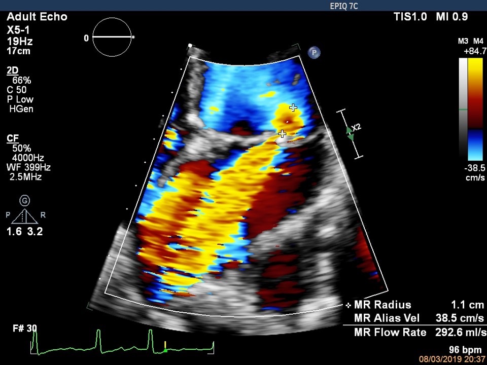
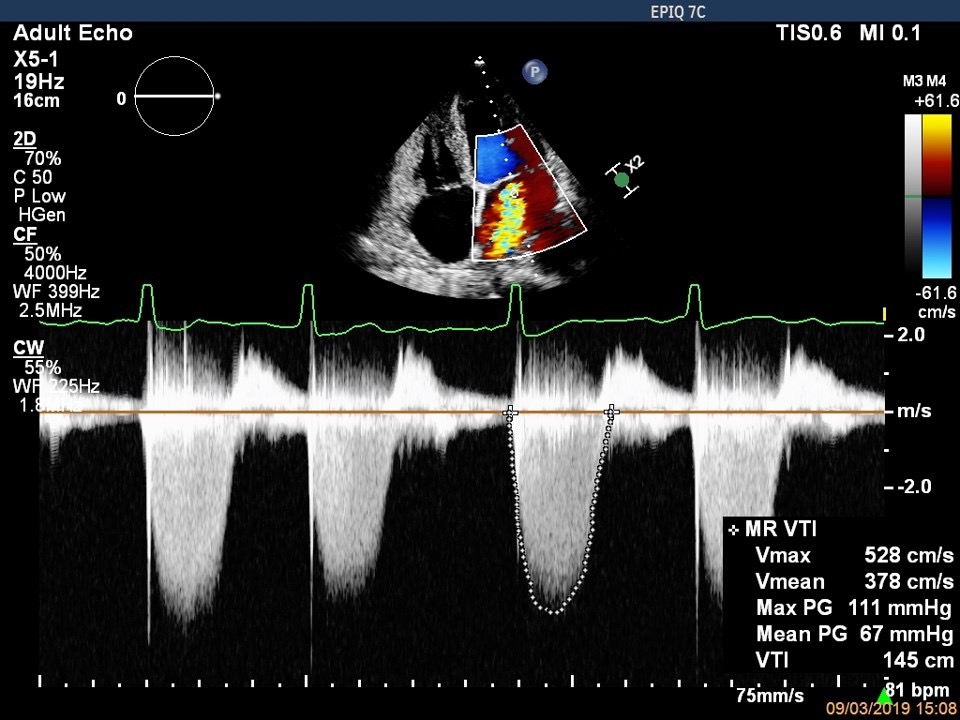
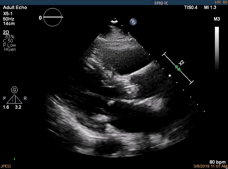
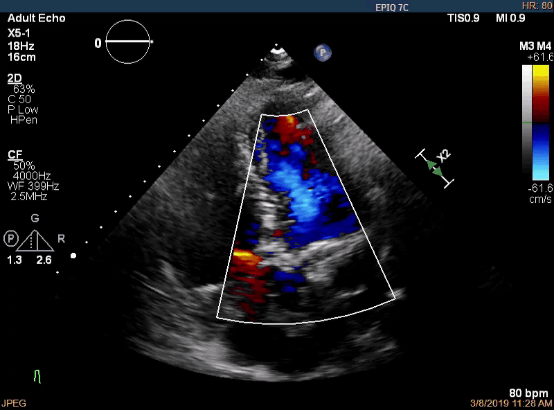
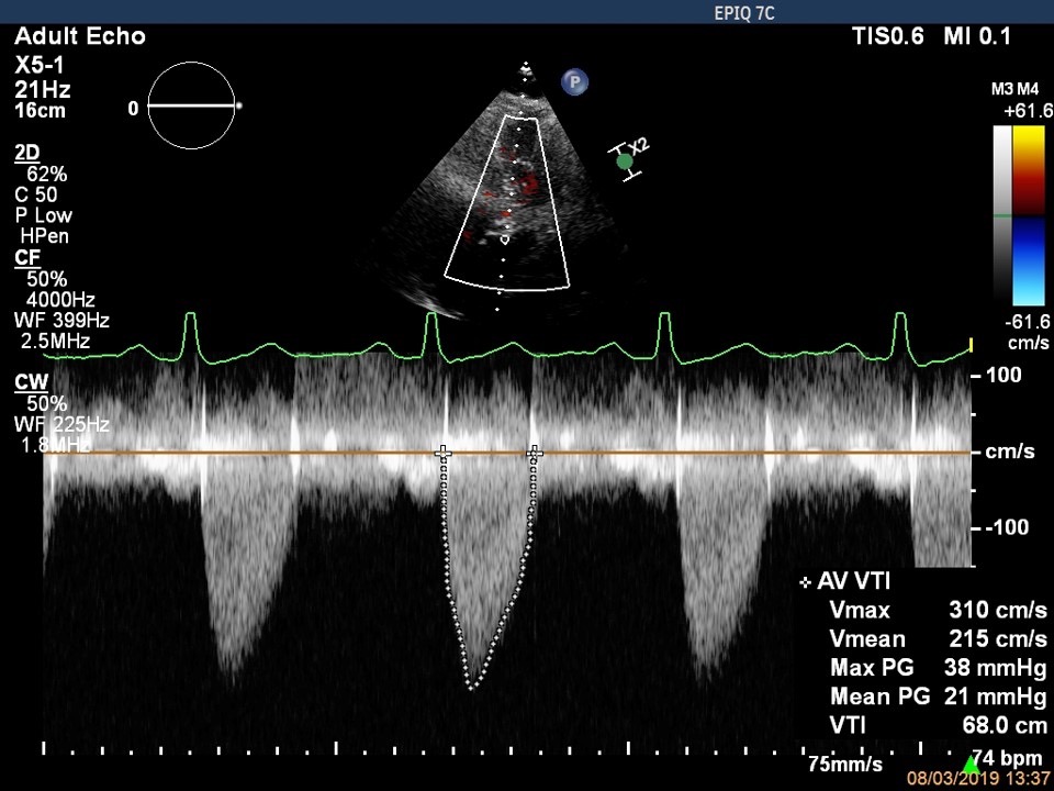
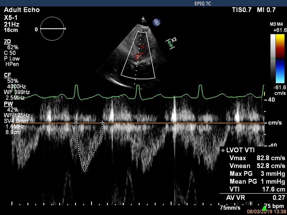
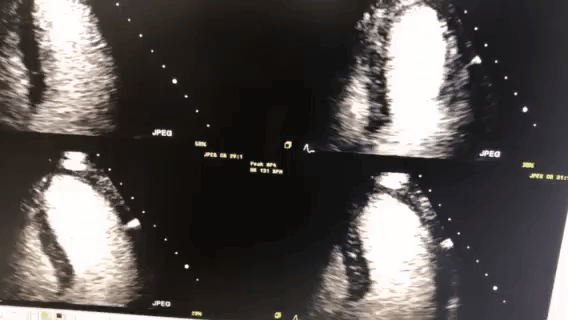
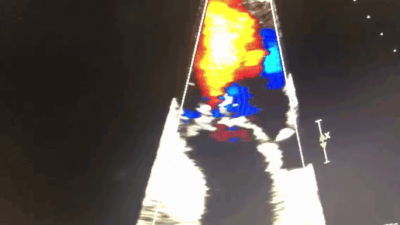
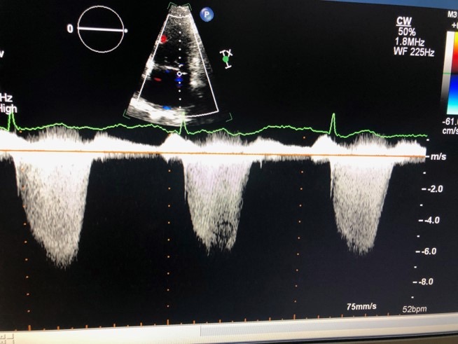
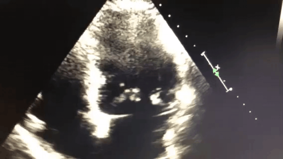
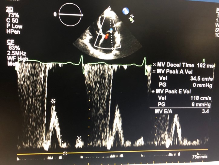
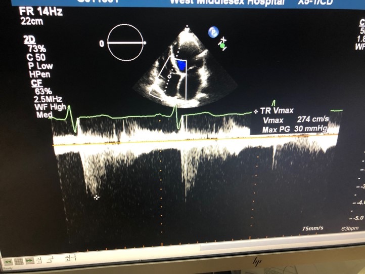
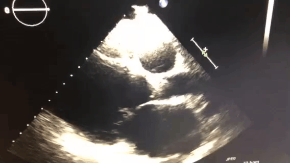
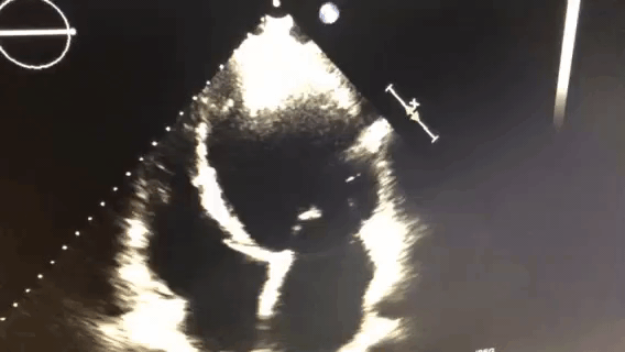
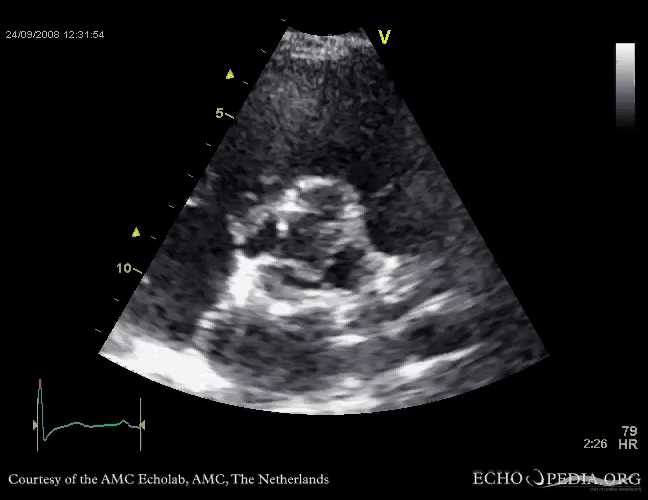
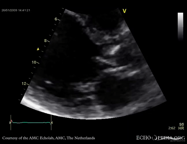
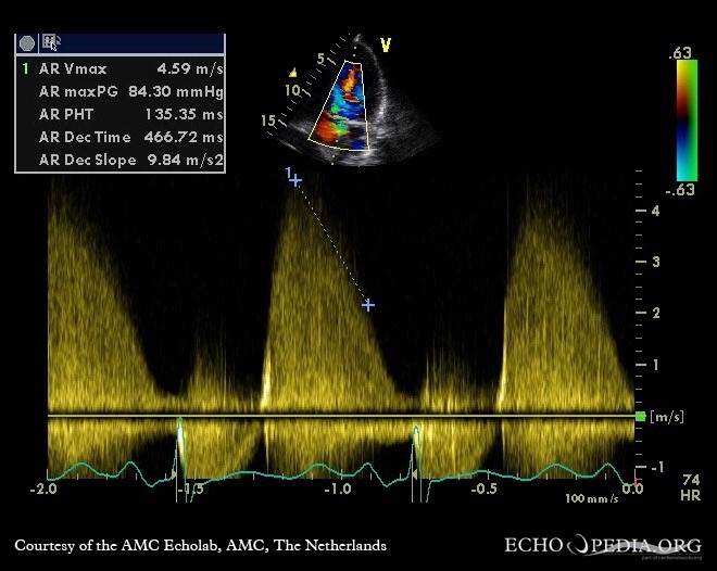
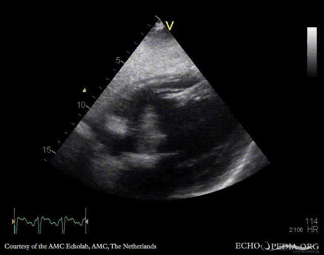
# Case 1 (Q22-Q27)
72F presents with breathlessness and peripheral oedema.
ECG: Atrial fibrillation
CXR: pulmonary oedema.
![]()
![]()
![]()
![]()
The LV function is:
- > 55%
- 50-55%
- 30-35%
- 40-45%
- 45-50%
The MR is:
- Anteriorly directed
- Posteriorly directed
The vena contracta of 0.92 suggests:
- Mild MR
- Moderate MR
- Severe MR
- Not enough information
The PISA radius suggests:
- Mild MR
- Moderate MR
- Severe MR
- Not enough information
Based on the data what is the regurgitant flow?
- 408ml/s
- 330ml/s
- 380ml/s
- 420ml/s
- 300ml/s
What is the EROA?
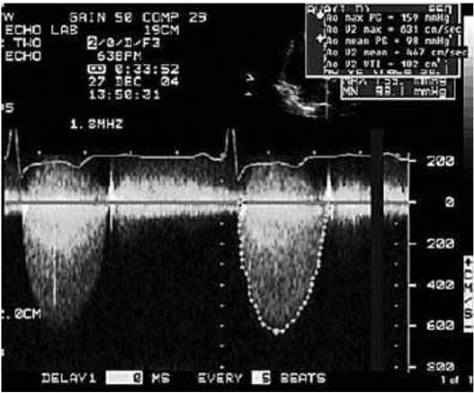
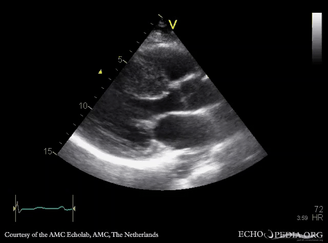
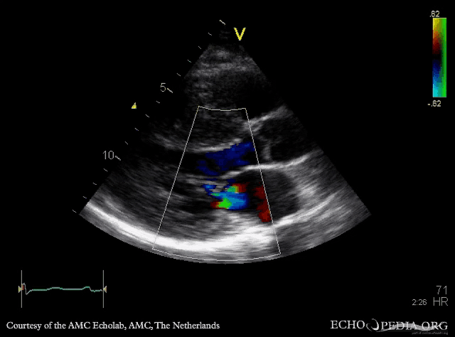
# Case 2(Q28-Q30)
73 year old female with chest pain
ECG: LVH
CXR: unremarkable
![]()
![]()
![]()
![]()
What is the left ventricular function?
- > 55%
- 45-50%
- 35-40%
- 30-35%
- < 20%
This patient has:
- Mild aortic stenosis
- Severe aortic stenosis
- Moderate aortic stenosis
- Aortic sclerosis
C
The patient has a dimensionless index of:
B
# Case 3 (Q31-Q32)
48 year old male with breathlessness.
ECG: normal sinus rhythm
CXR: unremarkable


![]()
What is the right atrial pressure?
- 5-10mmHg
- 0-5mmHg
- 15-20mmHg
- 30-35 mmHg
The PASP is:
- 46 – 51mmHg
- 64 – 69mmHg
- 54 – 59mmHg
- 44 – 49mmHg
- Unable to calculate based on the data
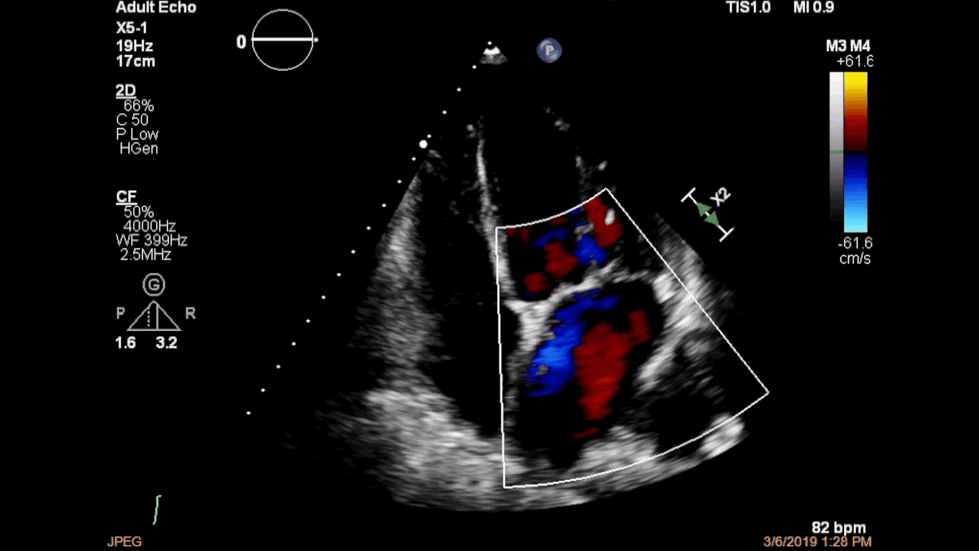
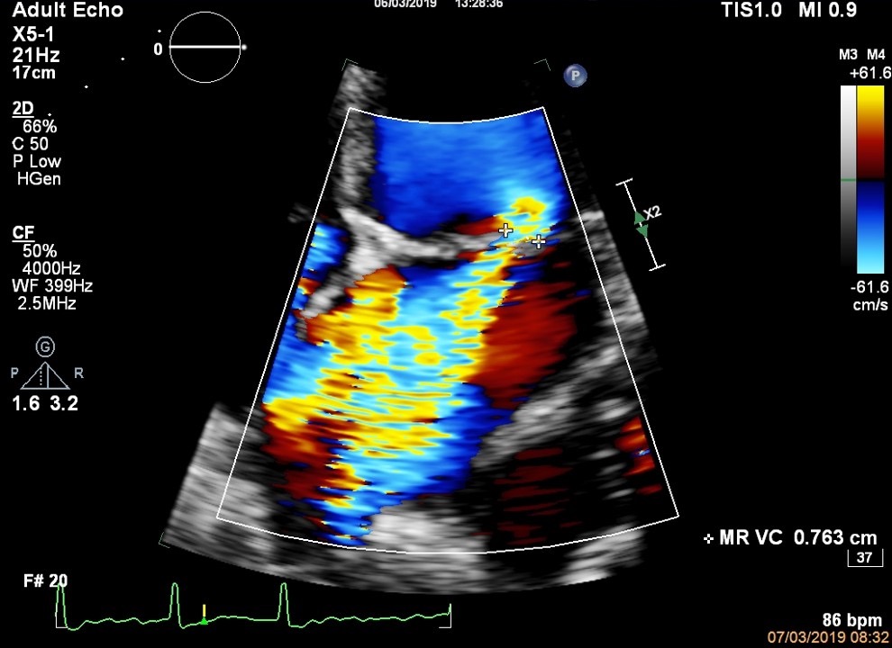
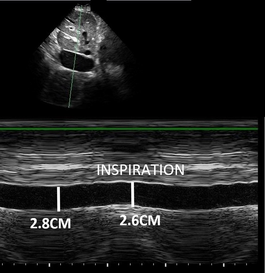
# Case 4 (Q33-Q34)
72 year old male with breathlessness and syncope.
ECG: LVH
CXR: unremarkable
![]()
![]()
This patient has evidence of (Which are true?)
- Asymmetrical LVH
- Symmetrical LVH
- Dilated left atrium
- Pericardia effusion
- Impaired ejection fraction
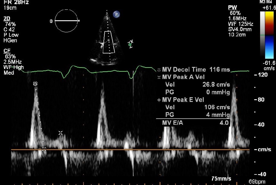
This MV inflow is suggestive of:
- Normal diastolic function
- Normal systolic function
- Mild systolic dysfunction
- Restrictive diastolic dysfunction
- Severe mitral regurgitation
# Case 5 (Q35-Q37)
32 year old male with a family history of sudden cardiac death.
ECG: anterior TWI
CXR: unremarkable
![]()
![]()
This patient has evidence of:
- Symmetrical HCM
- Sarcoid
- Amyloid
- Apical HCM
- Asymmetrical HCM
The mitral regurgitation appears:
- Mild
- Moderate to severe
- Severe
- No evidence of MR
The likely mechanism of the MR is:
- Infective endocarditis
- Flail anterior leaflet
- Systolic anterior motion mitral leaflet
- Prolapse mitral leaflet
- Restrictive mitral valve leaflet
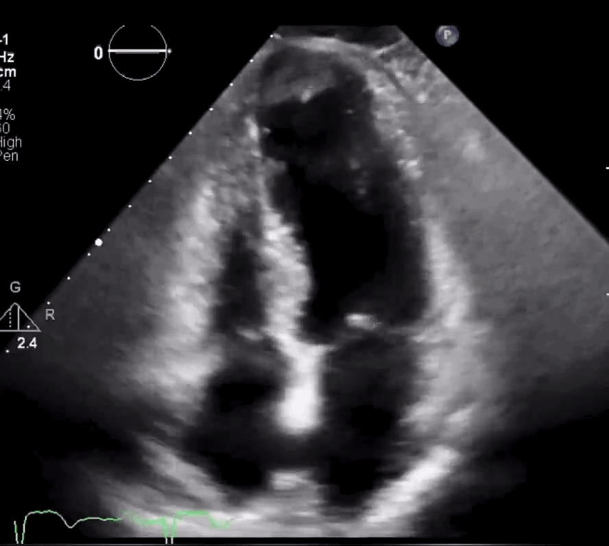
# Case 6 (Q38-Q40)
72 year old female with breathlessness and a recent stroke.
ECG: anterior ST elevation
CXR: fluid overloaded
![]()
What is the ejection fraction?
What is the cause of the LV dysfunction?
- Takotsubo cardiomyopathy
- Hypertrophic cardiomyopathy
- Amyloidosis
- Sarcoidosis
- Fabry’s disease
The following may be found in severe pulmonary hypertension: (Which are true?)
- TR Vmax of 4m/s
- A fixed and dilated IVC
- Dilated right ventricle
# Case 7 (Q41-Q42)
![]()
Which statement is true:
- Stress echocardiogram; 4 chamber view
- Stress echocardiogram; 2 chamber view
- Transesophageal echo; 4 chamber view
- 4 different patients
Which statement is true?
- Septal hypokinesis
- Normal LV wall motion
- Lateral hypokinesis
- Dilated RV
# Case 8
![]()
Which statement is true?
- 4 chamber; Mitral Regurgitation
- 2 chamber; Mitral Regurgitation
- 3 chamber; Aortic Regurgitation
- 3 chamber; Mitral Regurgitation
- 4 chamber; Tricuspid Regurgitation
# Case 9
![]()
Which statement is true?
- Mitral valve; CW; mild MR
- Mitral valve; PW; mild MR
- LVOT; PW; mild HCM
- Mitral valve; CW; severe MR
- Aortic valve; CW; severe AS
# Case 10 (Q45-Q47)
![]()
Which statement is most likely true?
- This patient is likely to have mitral regurgitation 可能有二尖瓣反流
- This patient has evidence of pulmonary hypertension 有肺动脉高压的证据
- This patient has evidence of HCM
- This patient has evidence of aortic stenosis
- This patient has evidence of mitral stenosis
If this patient has mitral regurgitation, the most likely cause is:
- Flail mitral valve leaflet
- LV dilatation
- Mitral valve prolapse
- Endocarditis with mobile vegetation
- Unknown
These segments are visible (Which is False?)
- Apical Lateral
- Basal Lateral
- Basal Inferolateral
- Basal Inferoseptal
- Apical Septal
# Case 11
![]()
Comment on the transmittal flow
- Normal
- Mild diastolic dysfunction 轻度舒张功能障碍
- Moderate diastolic dysfunction 中度舒张功能障碍
- Severe diastolic dysfunction 重度舒张功能障碍
- Unable to comment as patient in atrial fibrillation 作为房颤患者无法评论
# Case 12 (Q49-Q50)
![]()
What is the estimated pulmonary artery pressure?
- 30 mmHg
- 40 mmHg
- 50 mmHg
- 60 mmHg
- Unable to comment
Does this patient have:
- Normal pulmonary arterial pressure
- Borderline pulmonary hypertension
- Moderate pulmonary hypertension
- Severe pulmonary hypertension
- Unable to comment
# Case 13 (Q51-Q53)
![]()
Which LV segment is not visible in this view?
- Basal inferolateral
- Basal anteroseptal
- Mid anteroseptal
- Apical inferolateral
- Mid anteroseptal
Which statement is true?
- Normal size LV; severe systolic dysfunction
- Dilated LV; mild systolic dysfunction
- Dilated LV; severe systolic dysfunction
- Normal size LV; moderate systolic dysfunction
- Dilated LV; normal systolic function
Which statement is most likely false?
- The anteroseptum is most likely scarred, non-viable
- The patient may have symptoms of heart failure
- The patient has MR from a flail mitral leaflet
- The patient may have dizziness
- The inferolateral wall is severely hypokinetic
# Case 14 (Q54-Q55)
![]()
Which statement is false?
- There is LV thrombus 左室血栓
- There is lateral wall hypokinesis 侧壁运动减退
- LV contrast administration may be useful 左室造影剂可能有用
- There is LV dilatation 左室扩张
- There is LA dilatation 左房扩张
Which statement is true?
- There is a pleural effusion 有胸腔积液
- There is a mitral valve vegetation 有二尖瓣赘生物
- The LV is normal 左室正常
- The inferior wall is akinetic 下壁无运动
- The IVS is hypokinetic 间隔运动减弱
# Case 15 (Q56-Q57)
![]()
Describe this aortic valve
- Normal
- Bicuspid
- It has a vegetation
- Quadricuspid
- Aortic stenosis
Which valves are visible?
- Aortic, mitral, pulmonary
- Mitral, aortic, tricuspid
- Tricuspid, aortic, pulmonary
- Tricuspid, pulmonary, mitral
# Case 16
![]()
- Which statement is correct?
- There is aortic dissection 有主动脉夹层
- There is subaortic membrane
- There is aortic endocarditis
- There is mitral endocarditis
- Normal
# Case 17
![]()
Which statement is correct?
- CW; severe AS
- CW; severe AR
- PW; severe AR
- PW; severe AS
- CW; severe MR
# Case 18
![]()
Which statement is true?
- Pericardial effusion
- Mitral endocarditis
- Normal LV
- IVS rupture
- LV thrombus